Figure 1.
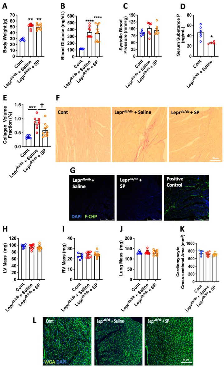
Leprdb/db mouse biometrics. (A) Body weight; (B) blood glucose; and (C) systolic blood pressure in control (Cont, n = 6), Leprdb/db + saline (n = 10), and Leprdb/db + SP mice (n = 10); (D) serum SP levels in Cont (n = 6) and Leprdb/db + saline mice (n = 4). Data are expressed as mean ± SD and were analyzed by one-way ANOVA with Tukey post hoc test, except for serum SP levels, which are expressed as mean ± SEM and were analyzed by unpaired t-test. * p < 0.05 vs. Cont, ** p < 0.01 vs. Cont, **** p < 0.0001 vs. Cont. Replacement SP ameliorates cardiac fibrosis. (E) Collagen volume fraction quantification; and (F) representative images of picrosirious red staining for Cont (n = 6), Leprdb/db + saline (n = 10), and Leprdb/db + SP mice (n = 10). Data are expressed as mean ± SEM and were analyzed by one-way ANOVA with Tukey post hoc test; *** p < 0.001 vs. Cont, † p < 0.05 vs. Leprdb/db + saline. (G) Representative images of unfolded collagen as identified by FAM-conjugated collagen hybridizing peptide (F-CHP) labeling of LV sections. Positive Control = LV section boiled in H2O to damage collagen prior to labeling with collagen hybridizing peptide (green fluorescence = unfolded collagen, Blue = DAPI). Replacement SP does not alter organ hypertrophy. (H) LV mass; (I) RV mass; and (J) Lung mass; (K) cardiomyocyte cross-sectional area; and (L) representative images of wheat germ agglutinin (WGA) staining for Cont (n = 6), Leprdb/db + saline (n = 10), and Leprdb/db + SP mice (n = 10). Data are expressed as mean ± SD and were analyzed by one-way ANOVA with Tukey post hoc test. (WGA = green, DAPI = blue).

