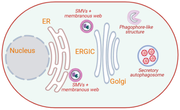
Figure 2.
Intracellular membrane rearrangements induced by picornaviruses. Picornaviruses, such as poliovirus and CVB3, induce the formation of SMVs that contain non-structural proteins and dsRNA, and are embedded in a membranous web located adjacent to the ER. Over the course of the infection, these membranous webs relocalize near the Golgi, where crescent-shaped phagophore-like structures emerge from them. These phagophore-like structures may serve as the precursors to double-membrane autophagosomes, which appear approximately 6 hr post-infection. Complete picornaviral particles appear to exit cells using secretory autophagy. (Image created in Biorender).

