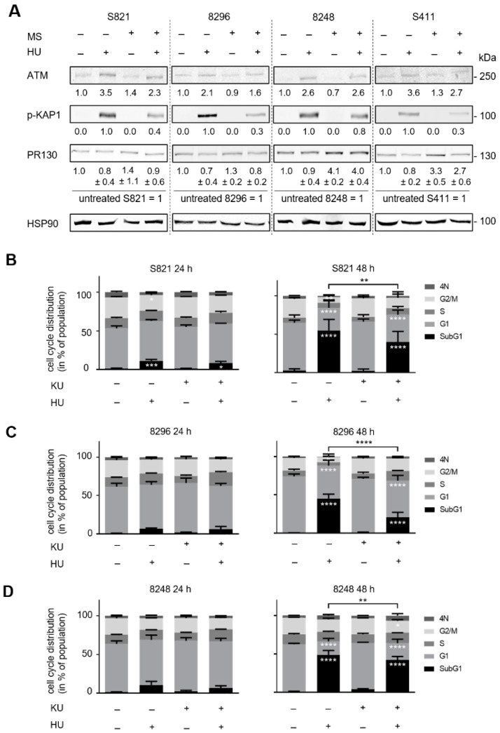
Figure 4.
Flow cytometry data showing the impact of hydroxyurea and KU-60019 on cell cycle progression and apoptosis-associated DNA fragmentation. (A) The PDAC cell lines S821, 8296, 8248, and S411 were treated with 5 µM entinostat ± 1mM hydroxyurea for 24 h. PR130 as well as the phosphorylation of KAP1 were measured by immunodetection; HSP90 as loading control; n = 3. (B–E) The cells were treated with 1 mM hydroxyurea (HU) ± 5 µM KU-60019 (KU) for 24 h and 48 h; n = 3. Flow cytometry was carried out to measure cell cycle distributions and subG1 phase cells. Statistical analysis was done with two-way ANOVA (* p < 0.05, ** p < 0.01, *** p < 0.001, **** p < 0.0001).


