Figure 5.
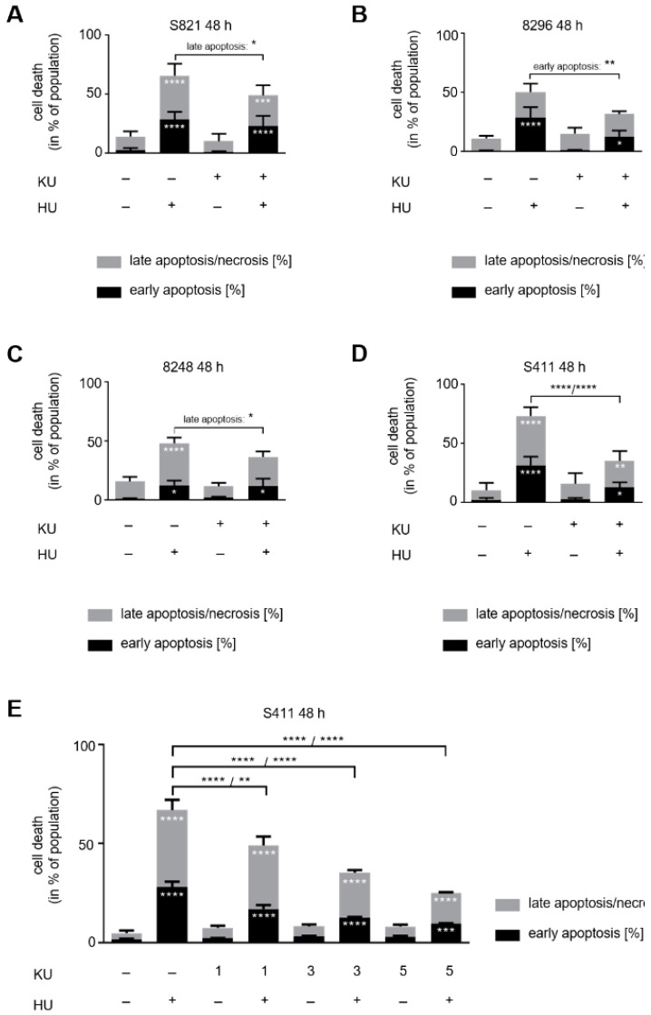
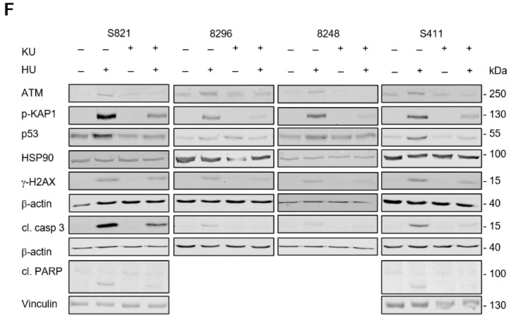
ATM inhibition counteracts apoptosis induction by hydroxyurea. Apoptosis analysis of (A) S821, (B) 8296, (C) 8248, and (D) S411 24 h after exposure to 1 mM hydroxyurea (HU) ± 5 µM KU-60019 (KU). Results were determined by flow cytometry using annexin-V-FITC staining and shown as mean ± SD (S821 n = 7; S411 n = 6; 8296/8248 n = 3). Statistical analysis was done with two-way ANOVA (* p < 0.05, ** p < 0.01, *** p < 0.001, **** p < 0.0001). (E) S411 cells were treated with 1 mM hydroxyurea and 1–5 µM of KU-60019. Results were collected by flow cytometry using annexin-V-FITC staining and are shown as mean ± SD (n = 3). (F) Immunoblot was done as indicated, with lysates from the four cell lines that were treated as mentioned above; fl., full-length PARP1; cf., cleaved form of PARP1; cl. casp. 3, cleaved form of caspase-3; HSP90, β-actin, vinculin as loading controls (S821 n = 3; 8296/8248/S411 n = 2).


