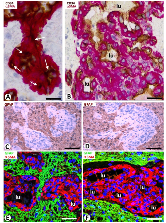
Figure 2.
Cellular components of the bizarre vessels and their relationship with neoplastic cells in GBM. (A,B) αSMA+ pericytes (red), increased in number and size, are observed around ECs (brown) of vessels with virtual lumen (A, arrows) or with small lumen (B, lu in a glomeruloid body). (C–F) The neoplastic glial cells (brown in C,D; green in E,F) are observed around glomeruloid bodies (hematoxylin-stained nuclei in (C,D); αSMA-stained pericytes in (E,F). Vessel lumen: lu). Note rows of neoplastic glial cells separating glomeruloid bodies in (C,E). (A,B) Double immunochemistry for CD34 (brown) and αSMA (red). Hematoxylin counterstain. (C,D) Immunochemistry for GFAP (glial fibrillary acidic protein) (brown). Hematoxylin counterstain. (E,F) Double immunofluorescence for GFAP (green) and αSMA (red). DAPI counterstain. Bar: (A,B) 20 µm, (C,D) 45 µm, and (E,F) 40 µm.

