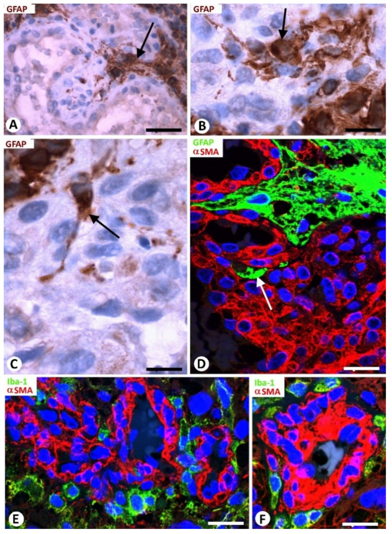
Figure 3.
Relationship of neoplastic glial cells (A–D) and microglia (E,F) with bizarre vessels in GBM. (A–D) Images of occasional neoplastic glial cells (brown in (A–C); green in (D), arrows) incorporated between glomeruloid body components (hematoxylin-stained nuclei in (A–C) and αSMA+ pericytes (red) in (D)). (E,F) Microgliocytes with an ameboid aspect (green) are observed around pericytes (red) of bizarre vessels. (A–C) Immunochemistry for GFAP (brown). Hematoxylin counterstain. (D) Double immunofluorescence for GFAP (green) and αSMA (red). DAPI counterstain. (E,F) Double immunofluorescence for Iba1 (green) and αSMA (red). Bar: (A) 40 µm, (B,C) 15 µm, and (E,F) 30 µm.

