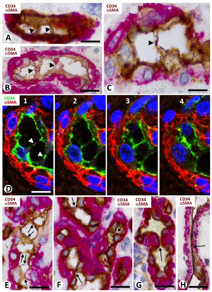
Figure 6.
Pillars and EC contacts in bizarre vessels of GBM. (A–E) EC bridges (arrowheads and arrows) are observed between opposite vessel walls. Note in (D1–4) the appearance and disappearance of pillars in a series of individual views in confocal microscopy. Two or more pillars can be observed in the same vessel (A,B,E). (F–H) Apical (F, arrows)) and planar (G,H, arrows) EC contacts from opposite vessel walls. Observe that the planar contacts can be with (G) or without (H) prominence of pericytes (red). (A–C,E–H) Double immunochemistry for CD34 (brown) and αSMA (red). Hematoxylin counterstain. (D1–4): Double immunofluorescence for CD34 (green) and αSMA (red). DAPI counterstain. Bar: (A,B,E–G) 20 µm, (C,D) 15 µm, and (H) 40 µm.

