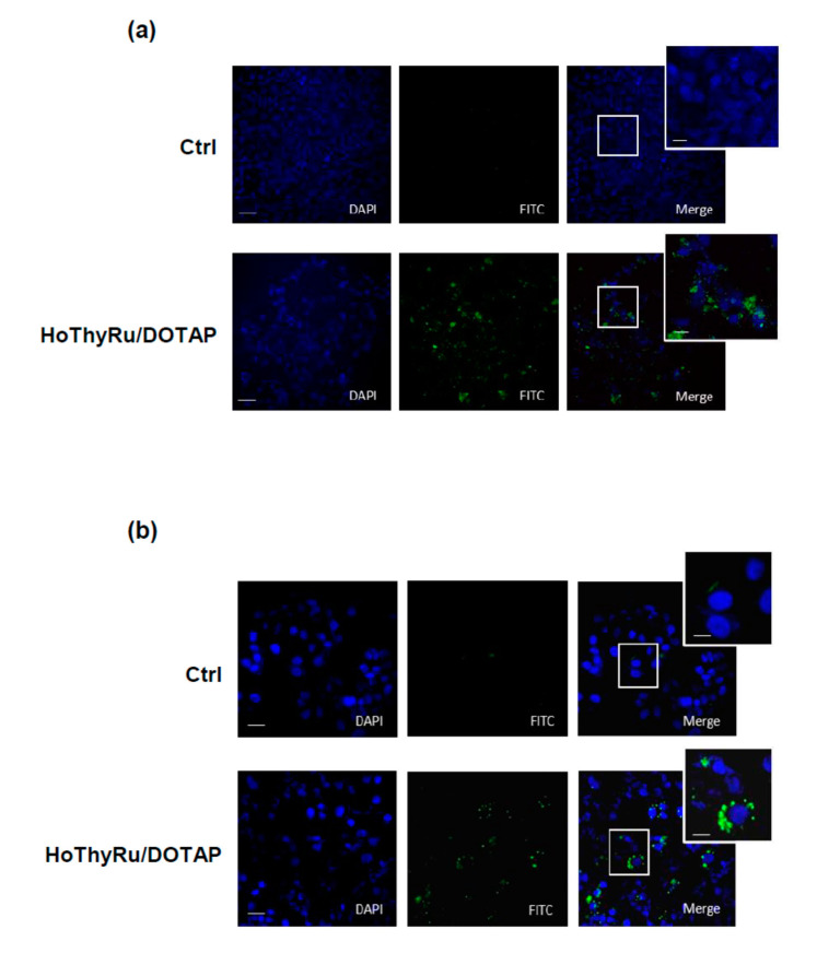
Figure A3.
(a) Apoptosis detection in MCF-7 cells following application in vitro for 48 h of IC50 of HoThyRu/DOTAP. A fluorescent “Apoptosis Detection kit” (Abcam, ab176749), designed to simultaneously monitor apoptotic and healthy cells, was used. The phosphatidylserine (PS) early apoptotic sensor is equipped with green fluorescence (Ex/Em = 490/525 nm, FITC filter); live cells are stained with a cytoplasm labelling dye (CytoCalcein Violet 450, Ex/Em = 405/450 nm, DAPI filter). MCF-7 were cultured in a black wall/clear bottom 96-well microplate and then treated or not for 48 h with IC50 of the HoThyRu/DOTAP nanoformulation. After incubation, cells were subjected to apoptosis detection according to the kit assay protocol. Fluorescence intensity was monitored by using a fluorescence microscope. (b) Autophagy flux detection in MCF-7 cells by a fluorescent Autophagic Detection Kit (Abcam, ab139484) following HoThyRu/DOTAP incubation in vitro for 48 h at the IC50 value. Nuclei are stained with blue nuclear stain (DAPI filter); autophagic vesicles (i.e., autophagosomes and autophagolysosomes) with green perinuclear and cytosolic stain (FITC filter). Experimental protocols, materials and methods are thoroughly described in [15] (Sci Rep. 2019; 9: 7006). In both (a,b), the fluorescent patterns from cell monolayers were overlapped to perform merged images (MERGE). The shown images are representative of three independent experiments.

