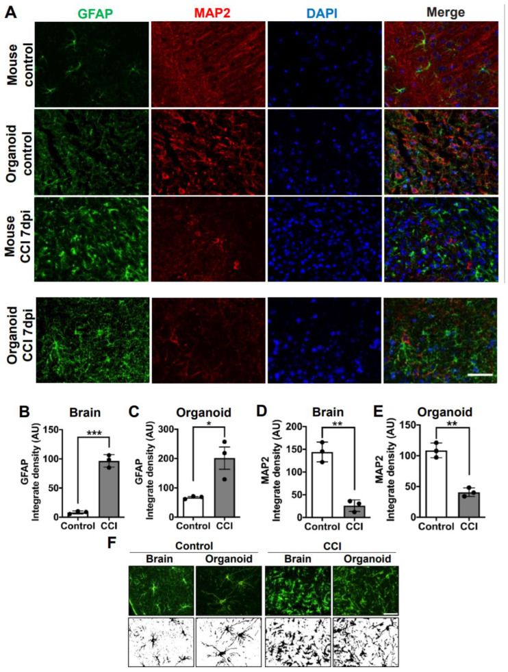
Figure 3.
Astrogliosis and reduction of neurons in COs after CCI. (A) Microphotographs of COs and mice brain subjected to CCI stained with GFAP and MAP2 antibodies to recognize astrocytes and neurons, respectively. Immunostaining was done 7 days after CCI. (B) Immunofluorescence quantifications of GFAP in mouse brain (Controls 8.241 ± 2.5 vs. CCI 96.68 ± 10.7; p = 0.0002) and (C) COs (Controls 67.31 ± 5.0 vs. CCI 201.6 ± 65; p = 0.0241). MAP2-positive neuronal density in (D) mouse brain (Control 144.2 ± 21.7 vs. CCI 26.24 ± 12.5; p = 0.0012) and in COs (E) (Control 108.7 ± 11.9 vs. CCI 40.73 ± 7.0; p = 0.001). (F) Morphological changes in astrocytes of COs and mouse brains were observed 7 days after CCI. Magnification: X40, scale bars = 50 μm. Statistical analysis performed with Student’s t-test, * p ≤ 0.05; ** p ≤ 0.01; *** p ≤ 0.001.

