FIG 1.

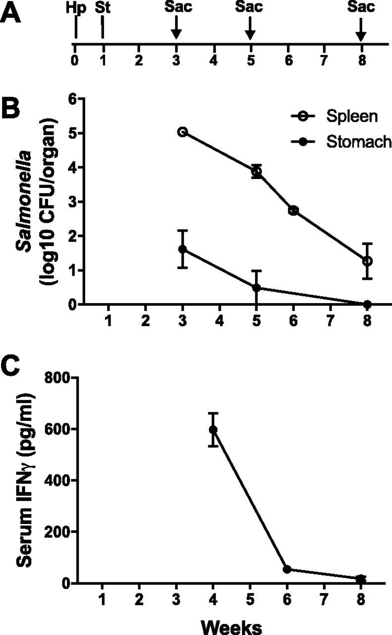
Characterization of the H. pylori-Salmonella coinfection model. (A) Schematic time frame of the H. pylori-Salmonella coinfection model. Mice were orally gavaged with H. pylori PMSS1 (Hp), infected intravenously with Salmonella Typhimurium (St) 1 week later, and sacrificed (Sac) 3, 5, or 8 weeks after H. pylori infection. (B) There was robust colonization of the spleen with Salmonella, which decreased over the course of infection. Salmonella was also present initially in the stomach at much lower quantities but was undetectable by 8 weeks. (C) Serum IFN-γ levels were high 3 weeks after Salmonella infection and declined rapidly. Data represent the means ± SEMs from 4 to 8 mice at each time point.
