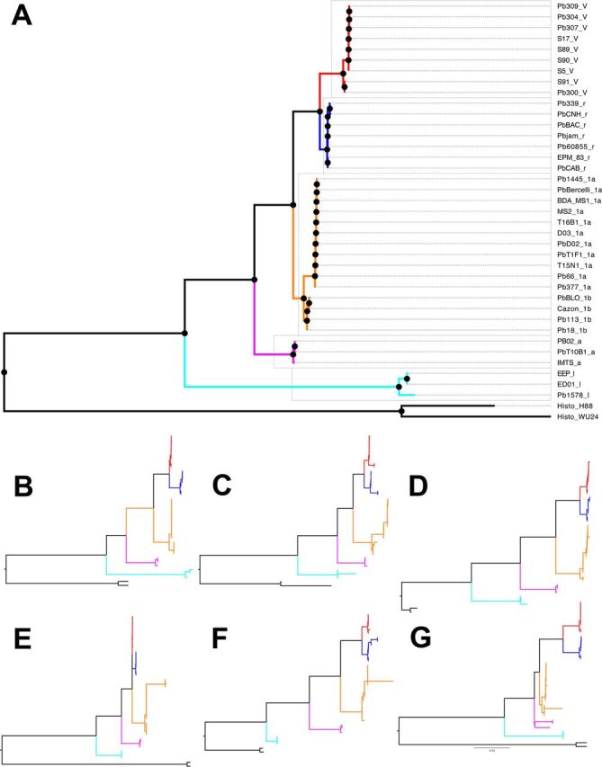
FIG 2.

Reciprocal monophyly between Paracoccidioides species. (A) Maximum likelihood rooted phylogram using concatenated genome-wide loci. Paracoccidioides venezuelensis isolates are marked in red and have a “V” after the strain name. Paracoccidioides restrepiensis isolates are marked in blue and have a “r” after the strain name. Paracoccidioides brasiliensis isolates are marked in orange and have a “1a” or a “1b” after the strain name. Paracoccidioides americana isolates are marked in pink and have an “a” after the strain name. Paracoccidioides lutzii are isolates marked in cyan and have a “l” after the strain name. (B to G) Phylograms for the six largest supercontigs in the Pb18 genome show the same topology. We follow the same color scheme as that in in panel A. (B) Supercontig 1.1. (C) Supercontig 1.2. (D) Supercontig 1.3. (E) Supercontig 1.4. (F) Supercontig 1.5. (G) Supercontig 1.6. The results are consistent to those shown in Fig. 1 of Muñoz et al. (13) and Teixeira et al. (25).
