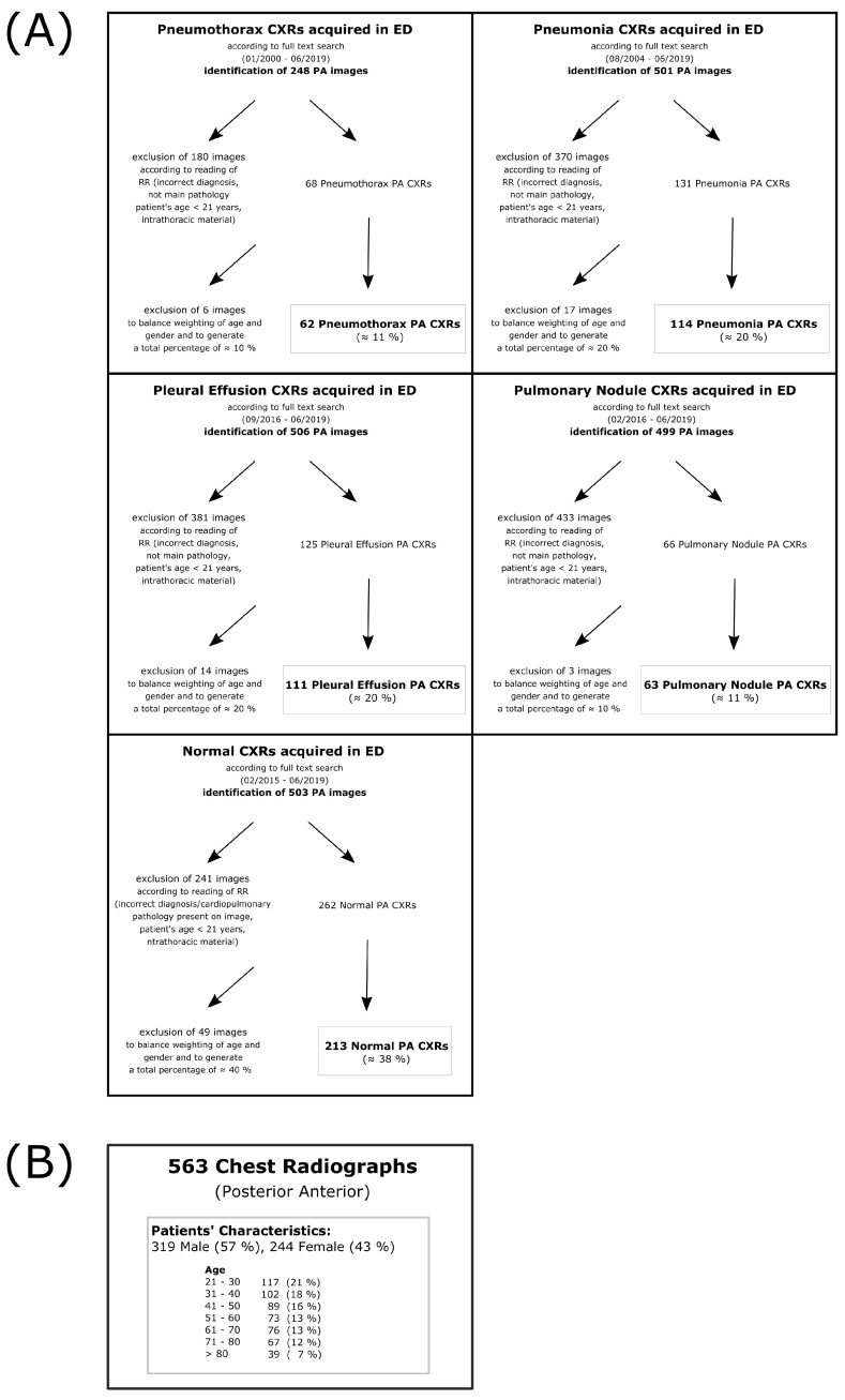
Figure 1.
Preselection of study cohort—(A) Flow charts that display the preselection process of each subcohort (normal, pneumothorax, pneumonia, pleural effusion and pulmonary nodule). Images were identified by full text search in the local PACS. All images were preread by a radiology resident not participating in the main reading process. Images that did not meet inclusion criteria (correct diagnosis, main pathology, patient’s age ≥ 21, no foreign material) were excluded. After a first preselection, further random images were excluded to balance out quantities in terms of age and gender in the different cohorts; (B) shows the overall patient’s characteristics in the final cohort. Notice that the preselection was based on the main pathology which means that also more than one pathology was possible (e.g., pleural effusion + basal consolidation or pneumothorax + pleural effusion). Frequencies could therefore also differ from board-certified radiologists’ evaluation (see Figure 2).

