Figure 1.
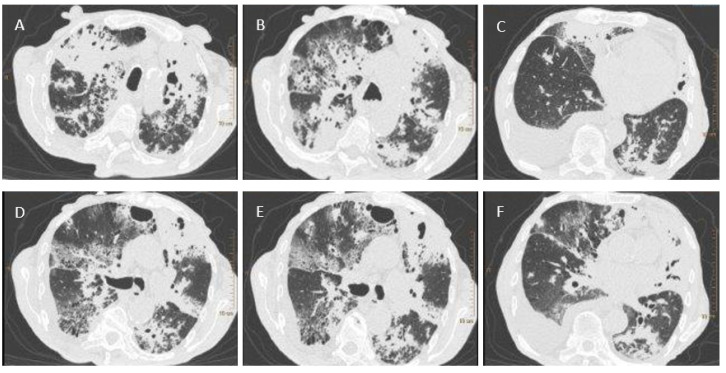
Figure 1. Chest CT scans. (A)—parenchymal-atelectatic lesions; thick-walled cavities filled with masses of tissue decay in the upper lobes; (B)—parenchymal-atelectatic lesions; cavities filled with tissue decay in segment 3 of the right and left lung, with the accompanying areas of tree-in-bud-appearance in segment 3 of the left lung and in the apex of segment 6 of the right and left lung; (C)—fluid in the pleural cavity; (D–F)—tree-in-bud-appearance in the apex of segment 6 of the right lung and in segments 6–9 of the left lung.

