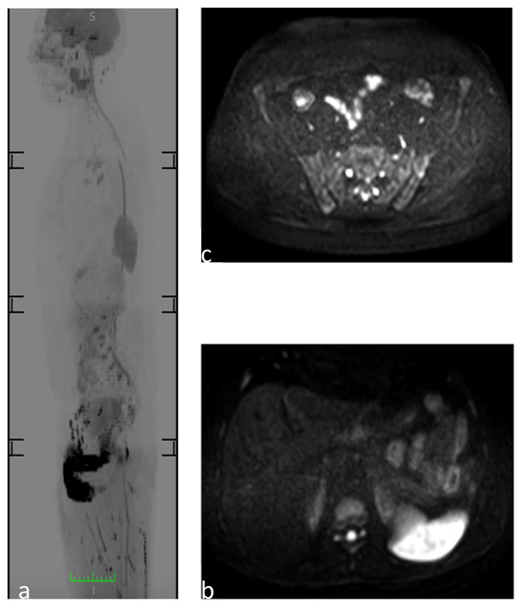
Figure 1.
WB-MRI to study a 30-year-old man. (a) Sagittal reformatted diffusion-weighted image MR images (inverted gray scale, high b values, b 1/41,000 s/mm2) showing a physiological restriction to diffusion of the spleen and genitourinary system. (b) Axial DWI image at the level of the upper abdomen showing the physiological restriction to diffusion of the spleen and the “t2 shine through artifact” of the medullary canal. (c) Axial DWI image at the level of the sacroiliac joint, which does not show the pathological areas of restriction to diffusion of the osteoarticular structures.

