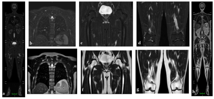
Figure 2.
WB-MRI to study a 30-year-old man (a). WB-MRI coronal STIR images do not show active inflammation consisting of synovitis or tenosynovitis, and pathological findings appear to be better visualized than in WB-MRI coronal T2 (h). (b–d) represent coronal STIR images, while (e–g) represent coronal T2 images at the level of the thoracolumbar spine; the hip and knees do not show the pathological hyperintensities or hypointensities of the signal in the osteoarticular structures under examination. (e) Hyperintensity in t2 at the level of the soma of a thoracic vertebra with isointenity in stir (b); findings compatible with angioma.

