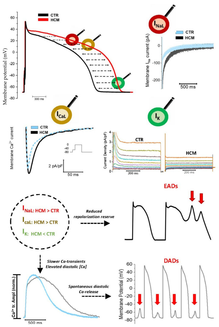
Figure 1.
Ion channel remodeling in HCM cardiomyocytes. APD is consistently prolonged in HCM myocytes, due to a combination of increased INaL, increased ICaL amplitude, slower ICaL inactivation and decreased potassium currents. APD prolongation raises the likelihood of EADs. Accumulation of intracellular calcium and Ca-overload is associated with enhanced spontaneous Ca-release from the SR, thus increasing the probability of calcium waves and DADs. Traces modified from Coppini et al. [26].

