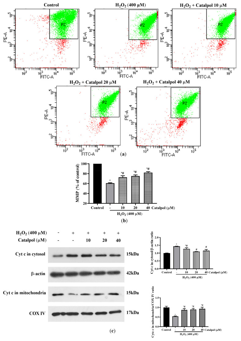
Figure 6.
Effect of catalpol on mitochondrial dysfunction in H2O2-treated RPE cells. (a and b) ARPE-19 cells were pretreated with or without catalpol (10, 20, and 40 μM) for 24 h and then treated with 400 μM H2O2 for 6 h. Changes in intracellular MMP were measured and analyzed with the JC-1 assay. The fluorescence intensity ratio of PE/FITC channels within the P2 gate was expressed as a change in MMP. In addition, to demonstrate the differences between the treatment and control groups in a more visual manner, we normalized the corresponding PE/FITC fluorescence intensity ratio of the treatment group relative to the control group. (c) Cytochrome c levels in the mitochondria and cytosol were measured using Western blot. COX IV was used as internal control of the mitochondrial fraction, and β-actin was used as internal control for the cytosolic fraction. Data are presented as mean ± S.D. of three independent experiments (* p < 0.05 vs. control group, # p < 0.05 vs. H2O2 (400 μM)-treated group).

