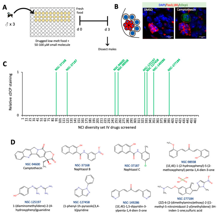
Figure 1.
Primary screen for GSC apoptosis in male fruit flies fed drug-spiked solid food (A) Diagram of experimental set up for NCI Diversity Set IV small molecule screen in low melt agarose food in male GSCs. (B) Diagram of male Drosophila GSC niche, wherein 6–12 GSCs (beige) are adjacent to niche cells (red), with all cells containing nuclei (blue). Representative images of germline from males treated with either vehicle control (DMSO) or camptothecin/NSC-94600. Stained with 1B1 (spectrosome, red), FasIII (hub, red), cDcp1 (apoptosis, green), and DAPI (nuclei, blue) (Scale bar 5 μm). (C) Visual representation of small molecules from the NCI Diversity Set IV which cause Dcp1 cleavage in male GSCs. (D) Chemical structures of small molecules that potentiated apoptosis in the male GSCs in the primary drug screen.

