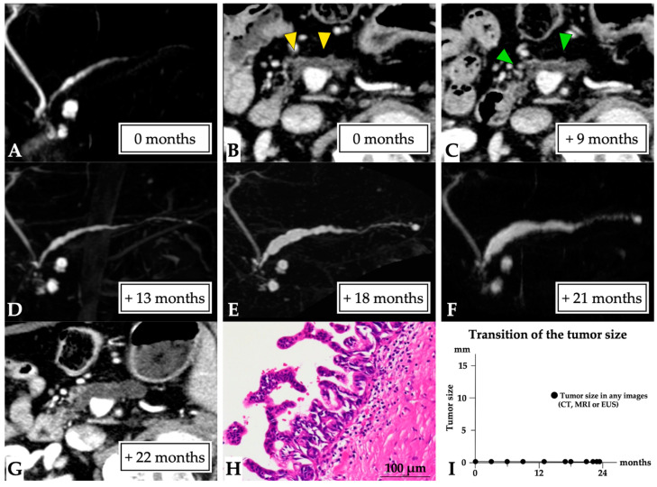
Figure 3.
Carcinoma in situ (0 mm in size) diagnosed over a 23-month observation period (Case 1 in Supplementary Table S1). A 79-year-old man was referred to our hospital for further examination concerning pancreatic head cysts that had been observed during abdominal ultrasonography screening. MRCP showed MPD stenosis in the pancreatic head with slight distal MPD dilation, in addition to the detected cysts ((A): first-time MRCP). CE-CT (B) and EUS images also revealed MPD dilation (yellow arrow head), but not a tumor lesion around the MPD stenosis; however, severe distal pancreatic parenchymal atrophy was detected. Therefore, these lesions were initially diagnosed as branch duct IPMN, and careful follow-up examination was required. Subsequently, although no changes to the pancreatic cysts and MPD abnormality findings were observed on CT (green arrow head: MPD dilation, (C)) and MRCP (D), MPD stenosis, and distal MPD dilation were clearly observed on MRCP after 18 (E) and 21 months (F). However, no tumor lesions were detected on CT (G) or EUS images. Endoscopic retrograde cholangiopancreatography was performed as malignancy was suspected. Pancreatic juice cytology was not performed because of failure to cannulate into the MPD. Despite the lack of a definitive diagnosis, the lesion was resected after 23 months because of possible malignancy. The final diagnosis was high-grade PanIN of the MPD, which had only spread in the MPD in the pancreatic head (Tis N0 M0, stage 0, final tumor size: 0 mm (CIS), (H)), along with retention cysts in the pancreatic head. This patient was diagnosed at 23 months after the first MRCP (I). Abbreviations: CIS, carcinoma in situ; EUS, endoscopic ultrasound; CE, contrast-enhanced; CT, computed tomography; IPMN, intraductal papillary mucinous neoplasm; MPD, main pancreatic duct; MRCP, magnetic resonance cholangiopancreatography; PanIN, pancreatic intraepithelial neoplasia.

