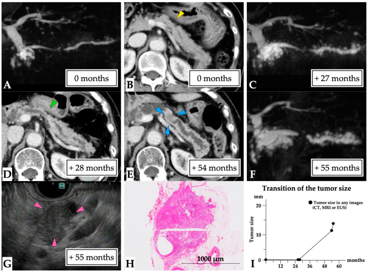
Figure 5.
A 14 mm lesion over a 55-month observation period (Case 5 in Supplementary Table S1). The case of a 75-year-old woman who had experienced idiopathic acute pancreatitis 7 months prior is presented. MRCP findings revealed MPD stenosis in the pancreatic body accompanied by distal MPD dilation and multilocular cysts in the pancreatic head (first-time MRCP, (A)). CE-CT scan images failed to detect a tumor lesion, although partial parenchymal atrophy consistent with MPD stenosis was observed (yellow arrow head, (B)); therefore, MPD stenosis was treated as chronic pancreatitis despite the absence of pancreatic stones. MRCP performed 27 months later showed progression of distal MPD dilation (C), although no tumor lesion was observed on CE-CT scan images (green arrow head: partial parenchymal atrophy, (D)). Finally, CE-CT scan performed 54 months later indicated the presence of a tumor lesion (blue arrow head, (E)). In addition, MRCP detected further MPD dilation (F) and the patient was referred to our hospital. EUS scans performed 55 months later showed a 14 mm diameter tumor (pink arrow head, (G)). EUS-FNA was performed, and the pathological diagnosis was adenocarcinoma (final tumor size: 14 mm). Therefore, distal pancreatectomy was performed for PC post-NAC. The final diagnosis was the presence of a 12 mm invasive nodule in the pancreatic body (T1 N1 M0, stage IIB, (H)). This patient was diagnosed 55 months after the first MRCP (I). Abbreviations: EUS, endoscopic ultrasound; CE-CT; contrast-enhanced computed tomography; CT, computed tomography; FNA, fine needle aspiration; IPMN, intraductal papillary mucinous neoplasm; MPD, main pancreatic duct; MRCP, magnetic resonance cholangiopancreatography; MRI, magnetic resonance imaging; NAC, neoadjuvant chemotherapy; PC, pancreatic cancer.

