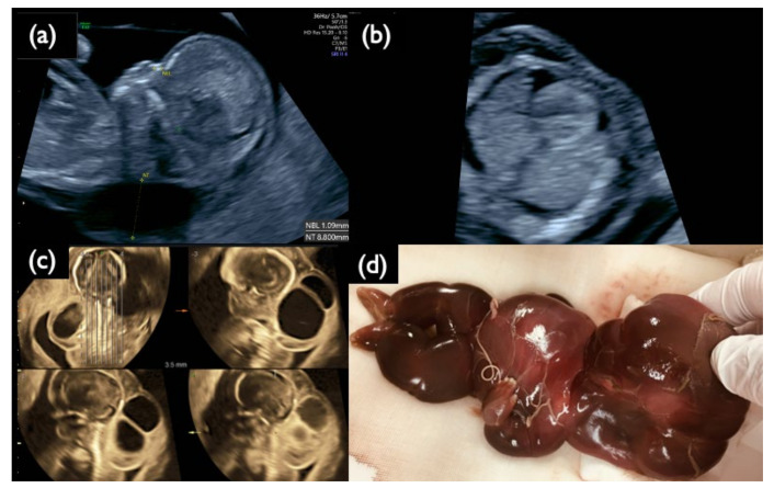
Figure 3.
Sonographic findings at 11+6 weeks, and 17 weeks, and IUFD fetus after delivery in Case FP1. (a) Mid-sagittal section of the fetal head. Nuchal translucency (NT) of 8.8 mm and a small nasal bone are demonstrated. (b) Horizontal section of the thoracic area. Pleural effusion is visualized bilaterally. (c) 3D tomographic ultrasound image in the sagittal section with a coronal guide section (left upper). General edema and large cystic hygromas are shown. (d) Picture of the dead fetus on delivery at 20 weeks. The fetus died in utero at 19 weeks of gestation.

