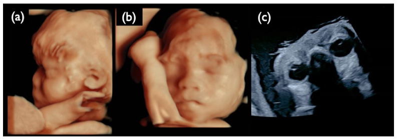
Figure 13.
Ultrasound images at 30 weeks of gestation in the NC1 case. (a) 3D ultrasound image of the fetal profile. (b) 3D ultrasound image of the frontal face of the fetus. (c) Ultrasound image of fetal eye sand lenses on both sides. All images show that the fetus is hypertrichotic and has no ocular lesions at this gestational age.

