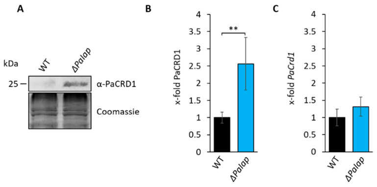
Figure 5.
Deletion of PaIap prevents proteolytic turnover of PaCRD1. (A): Western blot analysis of isolated mitochondria of 7-day-old wild type (WT) and ΔPaIap. Coomassie staining was used as loading control. (B): Quantification of PaCRD1 in (A). PaCRD1 levels were determined and related to wild type (set to 1). Data represent mean ± SD (n = 5). ** p < 0.01. (C): Transcript analysis of 5-day-old wild type and ΔPaIap grown on M2. PaCrd1 transcript level was analyzed by qPCR and normalized to PaPorin expression level. Data represent mean ± SD (wild type: n = 6, ΔPaIap: n = 3).

