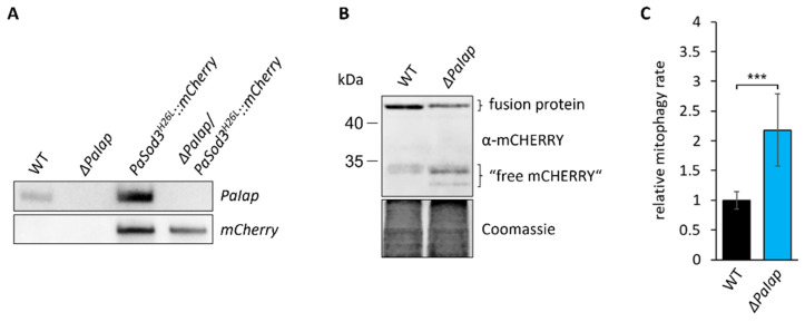
Figure 8.
Loss of PaIAP enhances mitochondrial turnover. (A): Southern blot analysis of HindIII-digested DNA of the generated mitophagy reporter strain ΔPaIap/PaSod3H26L::mCherry. (B): Western blot analysis of isolated protein extracts of 6-day-old PaSod3H26L::mCherry (here WT) and ΔPaIap/PaSod3H26L::mCherry (here ΔPaIap) grown in liquid CM medium. Coomassie staining was used as loading control. (C): Quantification of mitophagy rate in (B). Mitophagy rate was determined by ratio of “free mCHERRY” to total “free mCHERRY” + PaSOD3H26L::mCHERRY (fusion protein). Data represent mean ± SD (PaSod3H26L::mCherry n = 9; ΔPaIap/PaSod3H26L::mCherry n = 10). *** p < 0.001.

