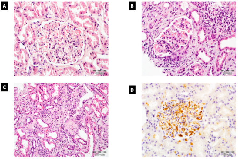
Figure 1.
Histopathology of IgAN. (A) Mesangial hypercellularity without endocapillary hypercellularity in glomerulus in IgAN; HE, 400×. (B) Glomerulus with cellular crescent and endocapillary hypercellularity. Presence of moderate interstitial inflammation; HE, 400×. (C) Extensive tubular atrophy and interstitial fibrosis in IgAN patient; HE, 200×. (D) Enhanced IgA reactivity in glomerulus (evaluated by immunohistochemistry); 400×.

