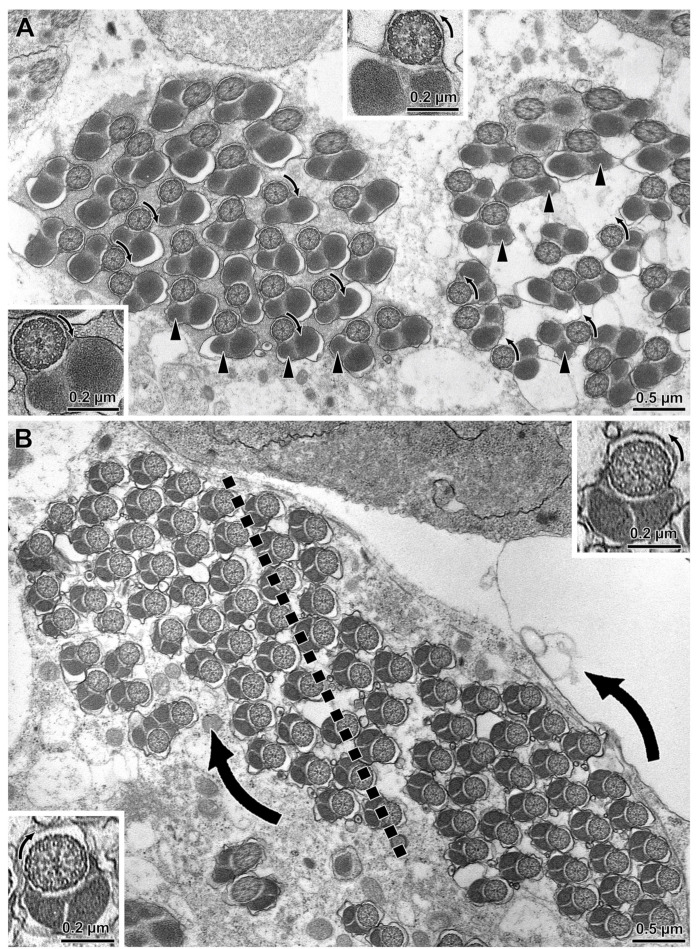
Figure 2.
Cross section through two adjacent loops of the same cyst of (A) D. willistoni and (B) D. saltans illustrating the bidirectional orientation of axonemes between loops. Note the position of the smaller mitochondrial derivative (arrowheads) differs between loops in being on the left or on the right side. In the loop to the left of both images, the dynein arms of the flagellar axonemes are oriented clockwise, whereas in the loop to the right the dynein arms are oriented anti-clockwise (arrows). The former (clockwise) are flagella seen from the tail end towards the nuclear region, whereas the latter ones (anti-clockwise) are flagella seen from the nucleus towards the tail end. Insets are higher magnifications of the flagellar axonemes with different dynein orientation.

