Figure 3.
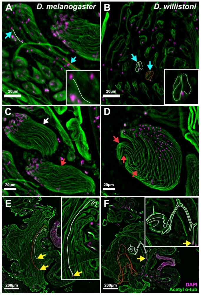
Epifluorescence micrographs of spermatid cysts from D. melanogaster (A,C,E) and D. willistoni (B,D,F) stained for acetylated α-tubulin (sperm tail, green) and DAPI (DNA, magenta). (A,B) Early stage of sperm tail elongation in a syncytial cyst of haploid spermatids. (A) D. melanogaster spermatid tails are straight (cyan arrows and trace lines). Both the cells and the sperm tails are elongated. (B) D. willistoni sperm tails are looped (cyan arrows and trace lines). The cells are still round, whereas the sperm tails have already elongated. (C,D) Slightly later stage of sperm tail elongation, showing groups of 64 spermatids encased in somatic cyst cells (cyst cells not shown). (C) D. melanogaster sperm tails are mostly straight (white arrow) with occasional kinks along their length (red arrow). (D) D. willistoni sperm tails are starting to bend and have multiple kinks along their length (red arrows). (E,F) Partially or fully elongated spermatid cysts. (E) D. melanogaster cysts are straight (trace lines). Note that the trace lines both start from a group of 64 sperm heads (yellow arrows) and extend along the entire length of each cyst. (F) D. willistoni cysts are looped multiple times (trace lines). Note that the white trace line covers the entire length of the cyst, starting at the 64 sperm heads (yellow arrows), but the red trace line covers only a portion of the cyst. For better visualization, images shown in (A,C) are maximum projections of z-stacks, whereas (B,D) are extended depth of field images processed using LAS-X software. (E,F) are single plane images. Insets are magnified 2.5 times compared to their corresponding images. Scale bars: 20 µm (A–D), 200 µm (E,F).

