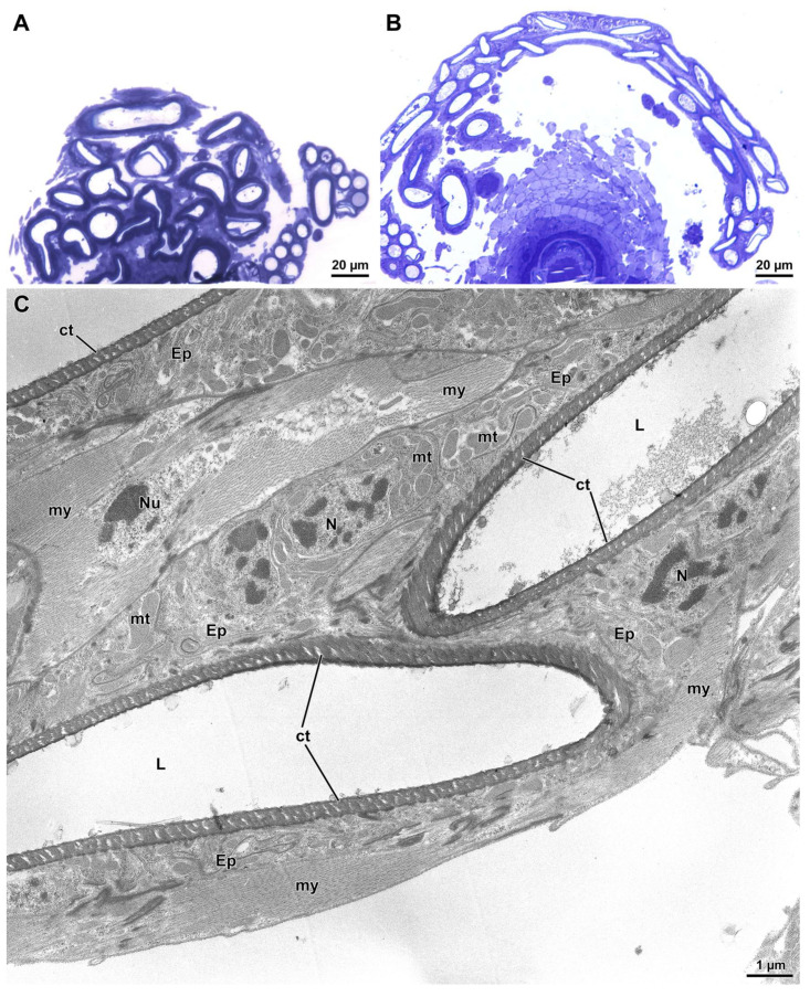
Figure 4.
The seminal receptacle (SR) of D. willistoni. (A) Semithin cross section of the SR at its insertion on the ventral region of the genital uterine wall. Note that the several cross-sectioned tubular loops are interconnected by a tissue complex. (B) Semithin section through a more distal region showing the integration of loops within a tissue complex. (C) Ultrathin section of the distal SR revealing that tubular lumens (L) are formed by an irregular thin epithelial layer (Ep) lined by a cuticle (ct). The cytoplasm contains nuclei (N), mitochondria (mt), and scanty dense inclusions. Beneath the epithelial layer, polymorphic muscle cells are visible with their nuclei (Nu) and the myofibrils (my).

