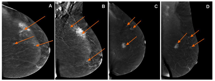
Figure 3.
Contrast-enhanced spectral mammography (CESM)subtraction images: (A) craniocaudal (CC) and (B) mediolateral oblique (MLO) views—irregular mass with heterogeneous enhancement and long spiculated mass BI-RADS 6. Numerous enhanced small foci are noted in left breast BI-RADS 4 (orange arrows); (C) craniocaudal (CC) and (D) mediolateral oblique (MLO) views—irregular mass with heterogeneous enhancement and long spiculated mass BI-RADS 6. Additional enhanced small foci are noted in the left breast in superior-outer quadrant BI-RADS 4 (orange arrows).

