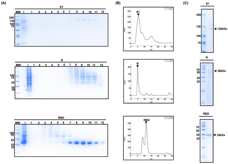
Figure 3.
Chromatography fraction analyses of the S1 domain of spike protein, nucleocapsid, and receptor-binding domain. (A) Coomassie-staining SDS-PAGE analysis of protein fractions obtained using fast protein liquid chromatography. After the proteins were eluted using a 500 mM Imidazole gradient, the fractions were subjected to purification using a Superdex 75 or Superdex 200 size-exclusion column, and elution was carried out using phosphate-buffered saline. Ten to fifteen fractions of 500 µL were obtained and then analyzed by sodium dodecyl polyacrylamide gel electrophoresis. Each lane represents a different fraction eluted. (B) Determination of the purity of eluted protein. The absorbance of the proteins contained in the eluted fractions was measured. Measurements were carried out at 280 nm and milli-absorbance units (mAU) were plotted (X-axis) against the elution volume (Y-axis). The peaks with the highest mAU values correspond to the fractions enriched with pure protein. (C) These fractions were analyzed by sodium dodecyl sulfate polyacrylamide gel electrophoresis to corroborate the purity of the protein.

