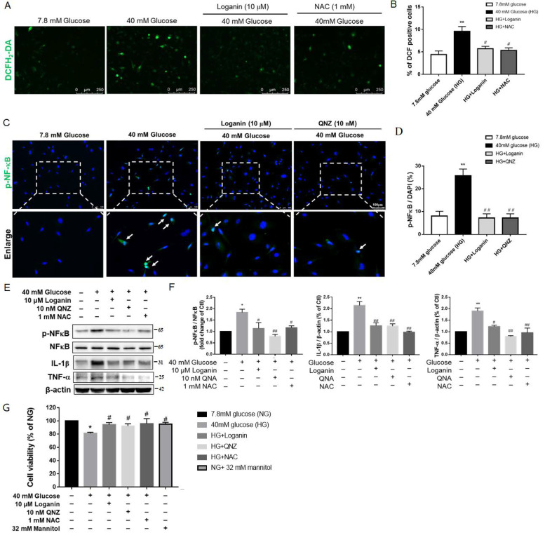
Figure 6.
Antioxidant and anti-inflammatory effects of loganin on high-glucose-treated SH-SY5Y cells. (A) Images represent intracellular ROS generation with DCF-positive cells in green (scale bar: 250 µm for magnification ×100) and (B) reflected as the mean gray value in each view (mean ± SEM, n = 10). Immunofluorescence staining revealed (C) phosphor-NF-κB (green) and DAPI (blue, nuclei) (scale bar:100 µm for magnification ×200). (D) mean gray value, respectively (n = 10 images from 4 groups, ** p < 0.01 vs. 7.8 mM glucose (NG); # p < 0.05 vs. 40 mM glucose (HG), ## p < 0.01 vs. HG). Western blot images show (E) the expression of p-NF-κB (Ser536), NF-κB, TNF-α and IL-1β proteins and (F) phosphorylation of NF-κB and fold changes of TNF-α and IL-1β proteins. β-actin was used as a loading control (n = 4). (G) Cell viability was measured by cell counting kit-8 (CCK-8). * p < 0.05 vs. NG, ** p < 0.01 vs. NG; # p < 0.05 vs. HG, ## p < 0.01 vs. HG. Data are expressed as mean ± SEM. QNZ: Quinazoline, NAC: N-acetylcysteine, ROS: reactive oxygen species, NG: normal glucose, HG: high glucose, NF-κB: nuclear factor-κB, TNF-α: tumor necrosis factor -α, IL-1β: interleukin-1β.

