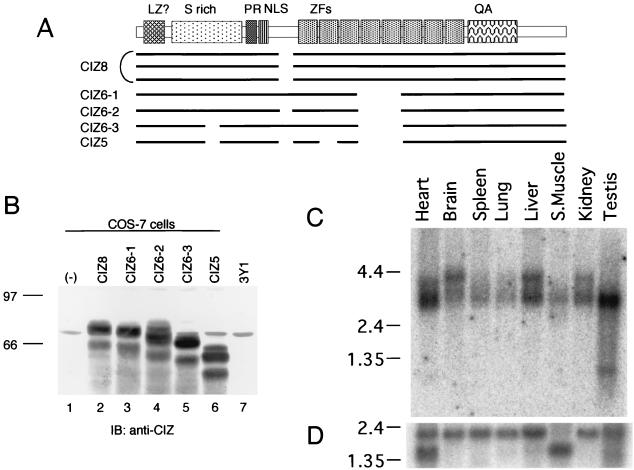
FIG. 2.
Alternative splicing forms and tissue distribution of CIZ. (A) Schematic diagram indicating the relationship among the clones obtained, including five alternative forms. LZ, leucine zipper; PR, proline rich; NLS, nuclear localization signal; ZFs, zinc fingers; QA, glutamine-alanine repeat. (B) Immunoblotting (IB) with anti-CIZ of the CIZ alternatives expressed in COS-7 cells and of the native CIZ in 3Y1 cells. The lanes are the lysates of mock (lane 1)-, CIZ8 (lane 2)-, CIZ6-1 (lane 3)-, CIZ6-2 (lane 4)-, CIZ6-3 (lane 5)-, and CIZ5 (lane 6)-transfected COS-7 cells and 3Y1 cell lysate (lane 7). (C) An RNA blot (Clontech) containing poly(A)+ RNA from the indicated rat tissues (2 μg/lane) was hybridized with a labeled CIZ partial cDNA probe (RsaI fragment corresponding to 234 to 481 bp). The positions of 9.5-, 7.5-, 4.4-, 2.4-, and 1.35-kb markers are shown on the left. (D) As a control, a blot with the cDNA of human β-actin is shown.

