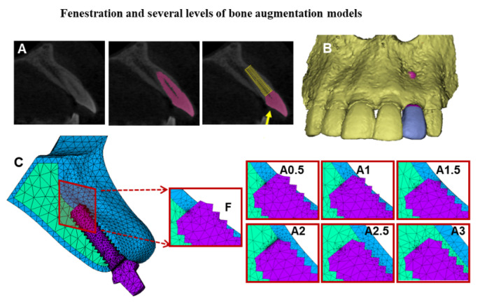
Figure 1.
The anterior maxillary bone segment with implant and abutment models. (A): The sagittal section of the lateral incisor tooth and implant inserted at the same direction of the tooth root; the yellow arrow indicates the applied force direction. (B): The three-dimensional CBCT based model of the anterior maxillary bone segment with facial bone fenestration defect. (C): Meshed fenestration model and simulation of several levels of bone augmentation of the facial bone defect are displayed in the cross-sectional images (F): Fenestration model, (A0.5–A3.0): Augmentation models with 0.5 mm intervals.

