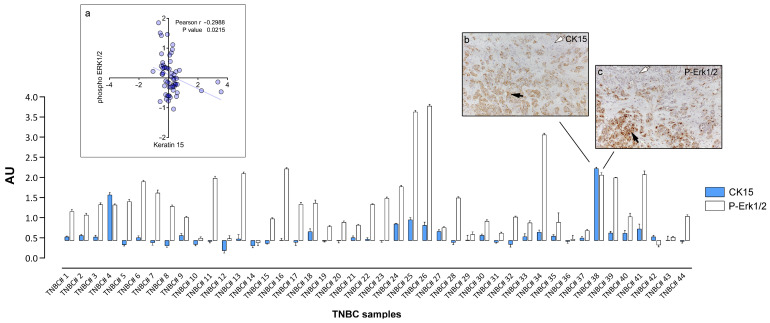
Figure 3.
Expression of CK15 and Erk1/2 phosphorylation in TNBC samples determined by IHC. Normalized RPPA values for CK15 and P-Erk1/2 were plotted using as baseline value the threshold RFI value of samples found negative by IHC analysis (subpanel a). IHC images of a heterogenous sample containing cancer cells positive (black arrow) and negative (white arrow) for CK15 (subpanel b) or P-Erk1/2 (subpanel c), respectively are presented for illustration purposes. Magnification, 20×.

