FIG. 3.
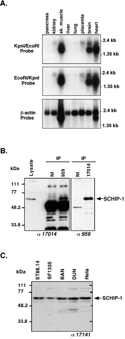
Expression pattern of SCHIP-1 mRNAs and proteins. (A) Northern blot analysis of SCHIP-1 expression. Human mRNAs from the indicated organs were analyzed by Northern blotting using either SCHIP-1 cDNA bp 1 to 405 (EcoRI/KpnI fragment), SCHIP-1 cDNA bp 402 to 2112 (KpnI/EcoRI fragment), or β-actin cDNA as the probe. Strong expression of SCHIP-1 is detected in skeletal (sk.) muscles, brain, and heart. (B) Immunoprecipitation of endogenous SCHIP-1 in ST88.14 cells. Endogenous SCHIP-1 from the schwannoma cell line ST88.14 was immunoprecipitated (IP) using either a chicken polyclonal antibody detecting a C-terminal region of SCHIP-1 (lane 959) or a polyclonal rabbit antibody detecting a central region of SCHIP-1 (lane 17014). Precipitates were resolved by SDS-PAGE on a 10% gel and detected with either rabbit antibody 17014 (left) or chicken antibody 959 (right). Twenty micrograms of crude protein extract from the ST88.14 cell line (Lysate) was loaded on the same gel. Antibodies used for Western blotting are indicated below the gels. NI, preimmune IgY or rabbit serum. The two independent antibodies 959 and 17014 both immunoprecipitate and detect in Western blots a protein migrating with an apparent molecular mass of 65 kDa. (C) SCHIP-1 expression in five human cell lines. Proteins (50 μg) from each cell line were resolved by SDS-PAGE on a 10% gel and detected with the rabbit polyclonal antibody 17141. Expression of the 65-kDa SCHIP-1 protein is detected in each cell line.

