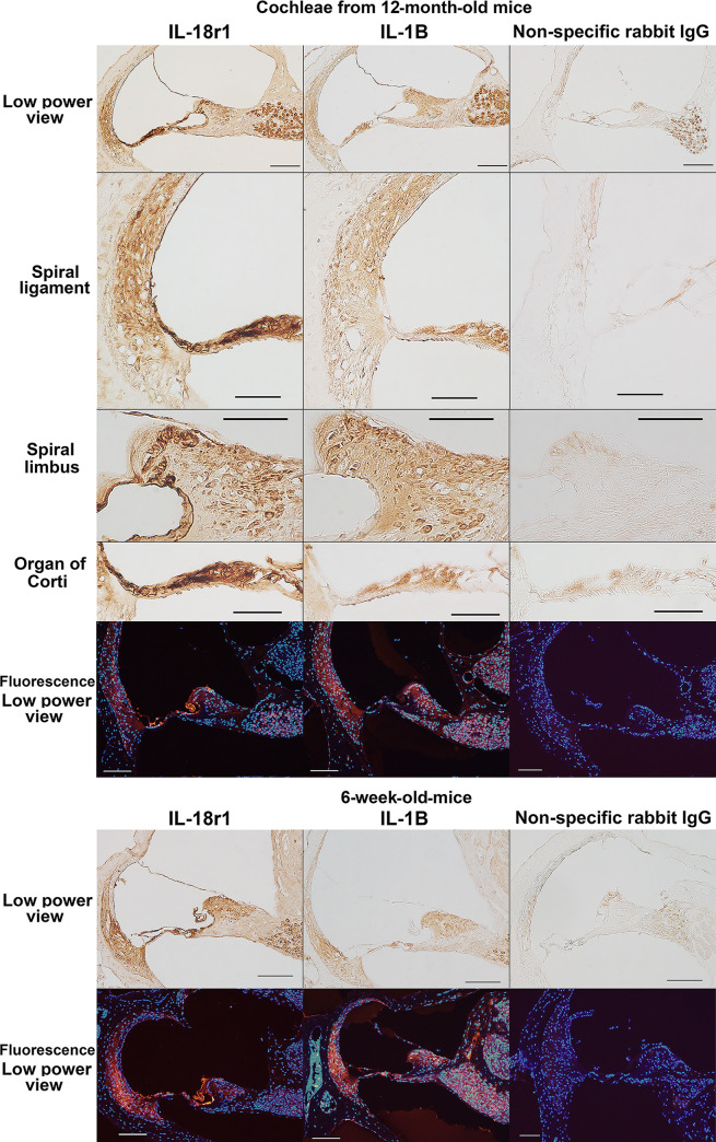
Fig 5. Immunohistochemical analysis of IL-18 receptor 1 (IL-18r1) and IL-1 beta (IL-1B) expression in aged cochleae.
The figures show immunoreactivity to IL-18r1 and IL-1B in the cochleae of 12-month-old and 6-week-old mice. Negative control sections incubated with nonspecific rabbit IgG showed no signals except for background staining in the spiral neurons in sections visualized using the ABC method. Therefore, the signals in the spiral neurons may be due to the nonspecific protein binding of rabbit IgG. In the immunofluorescent figures, blue and red indicate nuclear staining by DAPI and immunoreactivity to IL-18r1 and IL-1B, respectively. Scale bars indicate 100 μm (low-power view) and 50 μm (insets showing the spiral ligament, spiral limbus, and organ of Corti).

