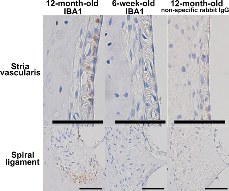
Fig 7. Immunohistochemistry showing IBA1-positive macrophages in aged cochleae.
The figures show immunohistochemical detection of IBA1-positive macrophages in the stria vascularis and the inferior division of the spiral ligament in the cochleae of 12-month-old mice. No IBA1-positive cells were observed in the cochlear structures of 6-week-old mice. Blue and brown indicate nuclear staining by hematoxylin and immunoreactivity to IBA1, respectively. Negative control sections incubated with nonspecific rabbit IgG showed no signals. Scale bars indicate 50 μm.

