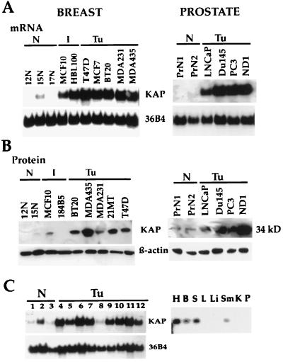
FIG. 1.
Overexpression of KAP in human mammary and prostate carcinoma cell lines. (A) Northern blot analysis showed overexpression of KAP mRNA in human mammary and prostate cancer cell lines (Tu) compared to normal human breast (12N, 15N, 17N) and prostate (PrN1, PrN2) epithelial cells. Twenty micrograms of total RNA, isolated from exponentially growing cells (70 to 80% confluence), was hybridized to a 32P-labeled KAP cDNA probe. A 36B4 cDNA probe was used as a loading control. I, immortal. (B) Western blot analysis was used to determine the level of KAP protein expression in several normal breast (12N and 15N) and prostate (PrN1 and PrN2) cell lines and in a number of immortal and cancerous mammary cell lines. Aliquots of cell lysates obtained from the indicated cell lines were loaded in each lane. Blots were probed with antibodies to KAP and β-actin. Immunoblotting for β-actin was used to achieve equal loading for protein samples. (C) KAP mRNA expression patterns in a variety of human normal and tumor cell lines (left) and in normal human tissues (right). Lanes 1 to 3, human normal cells; lanes 4 to 12, human cancer cell lines. Lane 1, hNMECs; lane 2, human normal urothelium; lane 3, human normal keratinocytes; lane 4, neuroblastoma cell line (SK-M-MC); lane 5, HeLa cells; lane 6, Jurkat cells; lane 7, 293 cells; lane 8, a renal carcinoma cell line (UMRC); lane 9, small-cell lung carcinoma cell line (H747); lane 10, melanoma (SK-MEL5); lane 11, osteosarcoma (HOS); lane 12, colon carcinoma cell line (HT29). Right, expression of KAP in human normal tissues. H, heart; B, brain; S, spleen; L, lung; Li, liver; Sm, skeletal muscle; K, kidney; P, pancreas.

