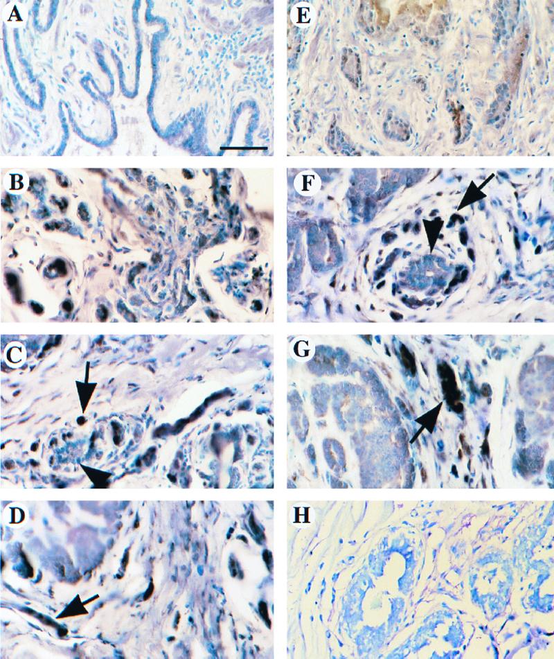
FIG. 2.
Tumor-specific overexpression of KAP in breast and prostate cancer patient samples. Expression of the KAP protein was evaluated using immunohistochemistry for a number of human normal and tumor tissue specimens of prostate (A to D) and breast (E to H). (A) Normal prostate gland had no noticeable staining of KAP protein. (B) Weakly invasive carcinoma of the prostate stained with high levels of KAP in epithelial cells. (C and D) Invasive carcinoma of the prostate showed intense cytoplasmic and nuclear immunoreactivity (arrow). Note normal prostate gland (arrowhead) with low or no KAP expression. (E) Normal breast epithelium had low expression of KAP. (F) Invasive carcinoma cells of the breast showed strong staining for KAP protein (arrow). Normal ducts with lower KAP expression are also shown in the same field (arrowhead). (G) KAP expression led to stronger staining in an invasive carcinoma than in a preinvasive ductal carcinoma in situ (arrow). (H) Negative control with normal immunoglobulin G showed no immunoreactivity in both invasive and ductal carcinomas in situ. Bar, 75 μm. Tissues were fixed in 3% paraformaldehyde and embedded in paraffin, and 5-μm-thick sections were processed for detection of the KAP protein. Immunocomplexes were identified by an ABC procedure, and 1% toluidine blue was used as a counterstain. There was little or no detectable expression of KAP either in the epithelial or stromal cells of ductal units of normal breast or prostate. However, in many tumors, large numbers of KAP proteins were displayed in a majority of cells.

