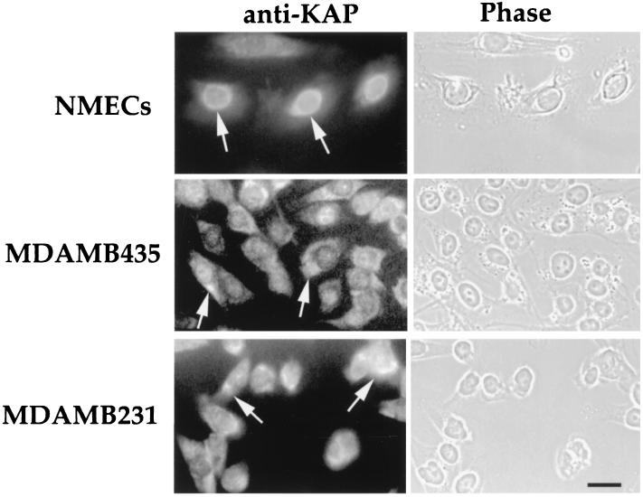
FIG. 3.
Subcellular localization of KAP in hNMECs (15N) and tumor mammary epithelial cells (MDAMB435 and MDAMB231). The cultures were fixed with 3.7% paraformaldehyde in PBS. Cells were permeabilized with 0.1% Triton X-100 prior to KAP antibody staining. Notice that the staining was restricted to the perinuclear region in hNMECs, while breast tumor cells (MDAMB435 and MDAMB231) had staining in both the cytoplasm and perinuclear area (arrows). The corresponding phase-contrast photograph is shown at the right of each fluorescence image. Bar, 10 μm.

