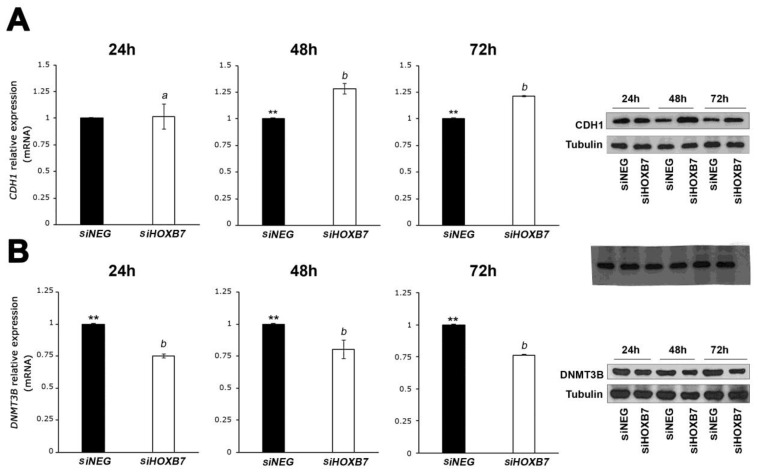
Figure 3.
Expression analyses of CDH1 (A) and DNMT3B (B) in MDA-MB-468 cells transfected with siHOXB7 and siNEG analyzed by RT-qPCR and western blots at three time points post-transfection. Three independent biological replicates were used in the statistical analyses comparing two variants (siNEG and siHOXB7) and the significant differences indicated with asterisks based on the analyses using t-test. The differences in the expression measures at the three time points were compared using the post hoc Tukey test. CDH1 expression was significantly higher in siHOXB7-transfected cells than in controls at 48 h and 72 h (**, p < 0.05). Comparing the three time points, the expression of CDH1 is significantly different at 24 h in these cells (a) in comparison with 48 h and 72 h (b). No significant differences in CDH1 expression were detected between 48 h and 72 h. DNMT3B expression was significantly lower in HOXB7-silenced cells than in the controls in the three time points analyzed, and no significant differences were detected in the expression between these time points (b). In the western-blot analyses, β-tubulin was used as the reference protein. a: significant expression differences between time-points in the siHOXB7 cells evaluated with post hoc Tukey test; b: no significant expression differences between time-points in the siHOXB7 cells evaluated with post hoc Tukey test.

