
Figure 2.
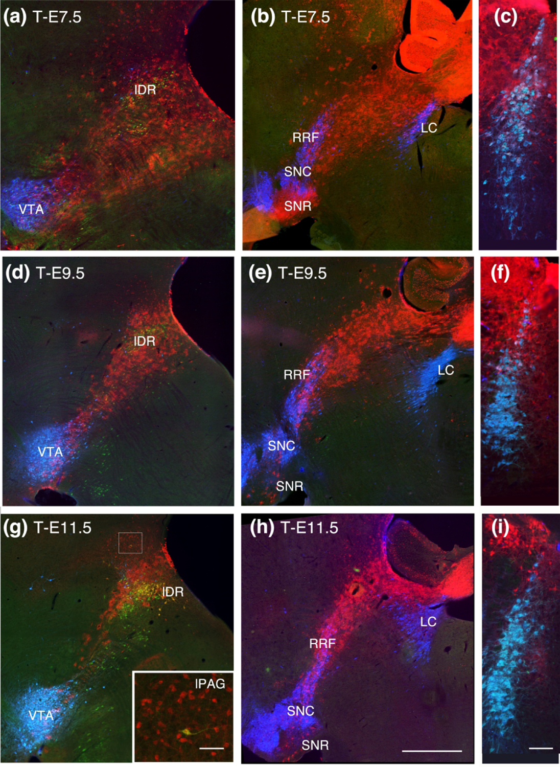
Sagittal sections progressively lateral to the midline, continuing the progression as shown in Fig 1. and coronal sections through the locus coeruleus (LC). (a, b, c) At T-E7.5 TDT surrounds serotonin neurons in the lateral wings of the DR (lDR) and extends caudally to include TH neurons in the rostral and dorsal LC. (d, e, f) Although there is considerable thinning of the TDT band at T-E9.5, the lateral DR and dorsal tip of the LC retain TDT labeling. (g) At T-E11.5 TDT remains dorsally located in lDR serotonin neurons and migrated neurons can be seen in the lateral PAG (inset in G). (h, i) The rostral extent of TDT is similar to previous timepoints and just dorsal to the LC. Sagittal sections show at the scale in (h), bar = 1 mm; inset in (g) bar = 100 um; (c, f, i) scale show in (i) bar = 200 um.