
Figure 4.
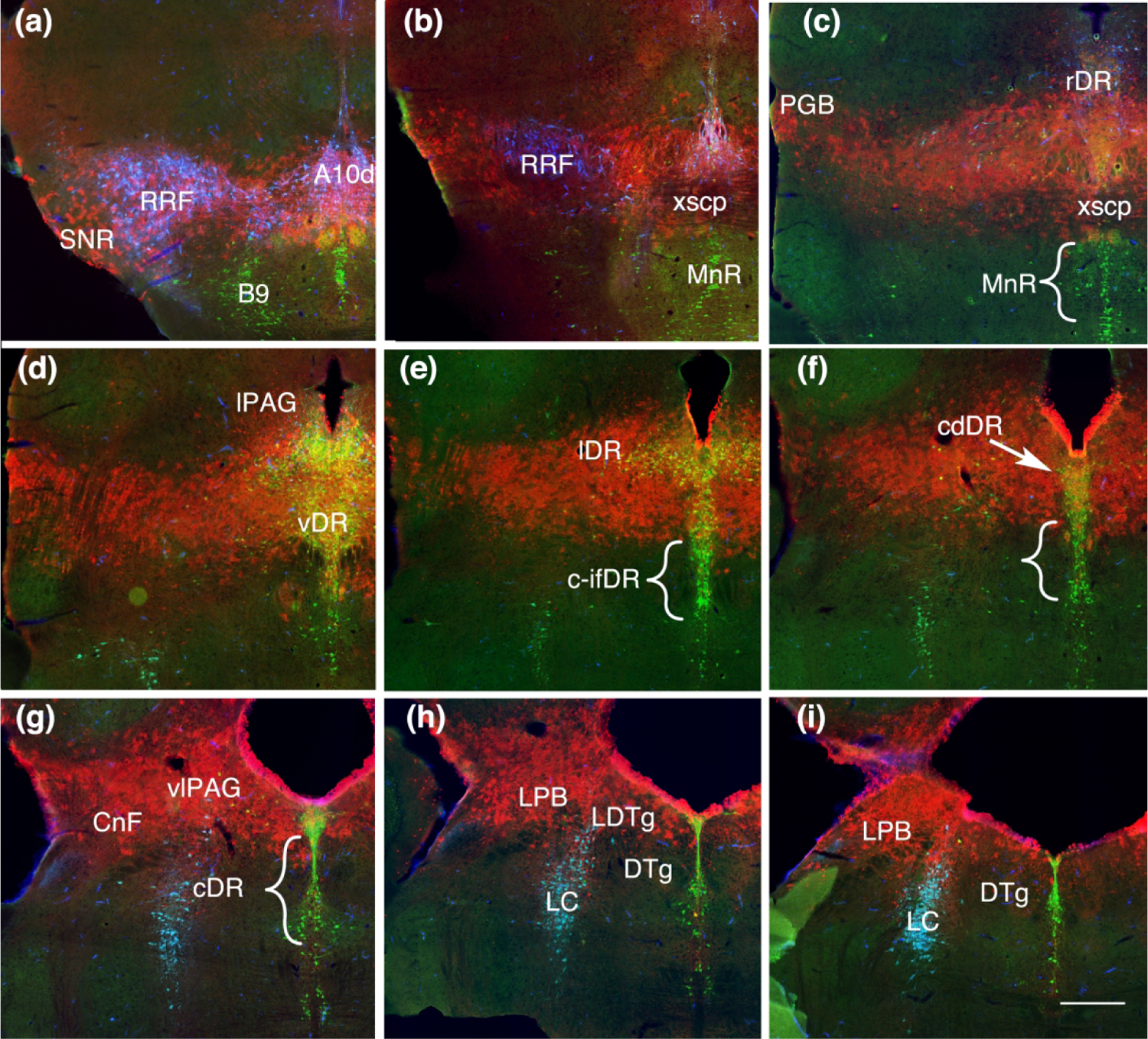
Coronal sections showing Fgf8 fate map at T-E9.5 (red, TDT) with immunolabeling for TH (blue) and TPH (green). (a) Rostrally TDT remains associated with the dopaminergic nuclei, but is not present within dopamine neurons. (b, c) TDT is within the xscp but not in serotonin neurons ventral to the xscp in the MnR. (d) All of the middle levels of the DR containing TDT. (e, i) Lateral wings (lDR) and caudal-dorsal DR (cdDR) contain TDT however serotonin neurons ventral in the caudal and caudal intrafasicular DR (c-DR c-ifDR) and areas medial to the DTg lack TDT. Scale bar in (i) = 250 um