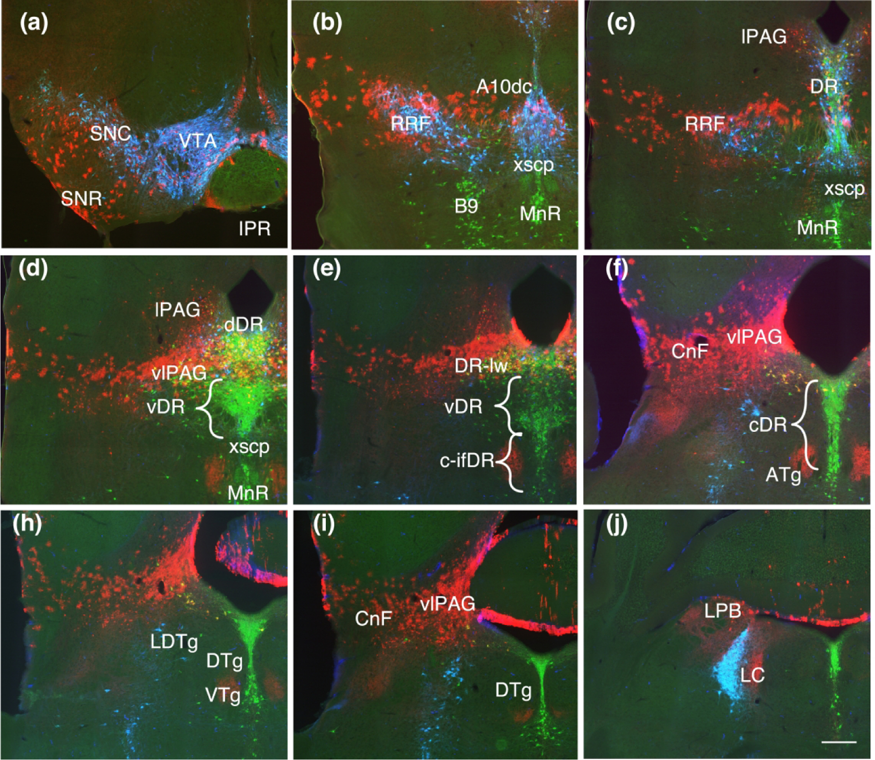
Figure 5.

Coronal sections showing Fgf8 fate map at T-E11.5 (red, TDT) with immunolabeling for TH (blue) and TPH (green). (a, b, c) TDT labeling in the dopaminergic nuclei follow the same pattern as with earlier timepoints but the band of labeling appears sparser and thinner, with a reduced ventral extent. (d) At this point, TDT is largely absent from the ventral DR (vDR) where serotonin neurons extend laterally in a triangular shape above the xscp. (e, f) Except for the caudal lateral DR (DR-lw) which retains TDT, the remaining caudal DR both dorsal and intrafasicular (c-DR, c-ifDR) is excluded from the TDT domain except for a few scattered neurons. (h, i, j) TDT labeling persists in the ventrolateral PAG (vlPAG) and cuneiform nucleus (CnF). Scale bar in (i) = 250 um
