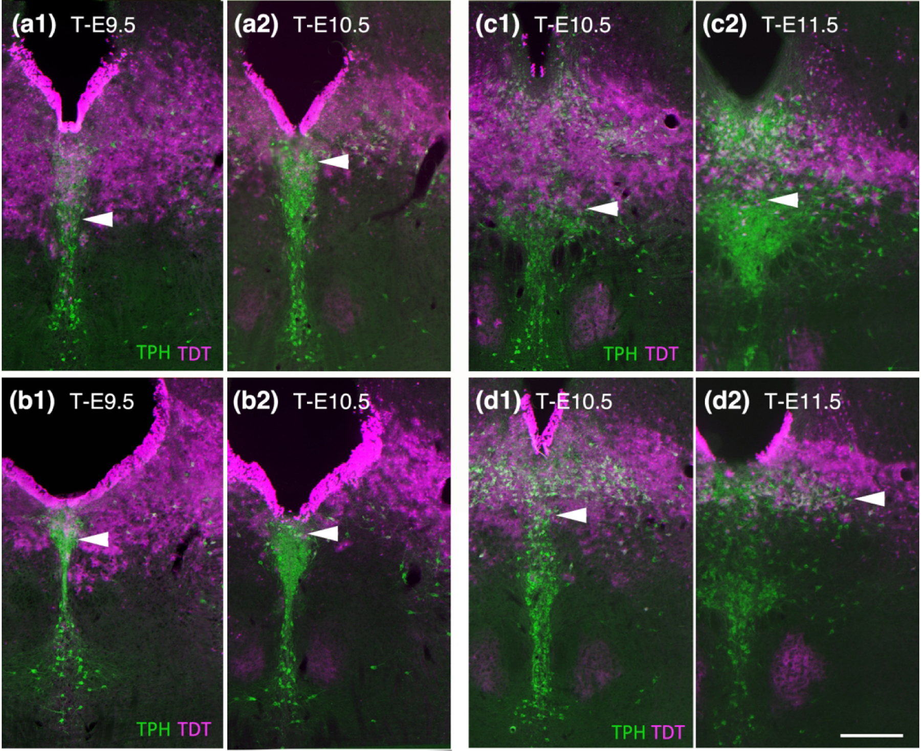
Figure 6.

A more detailed look at the ebbing of Fgf8 lineage through serotonin neurons in the DR comparing T-E9.5 to T-E10.5 (a, b) and T-E10.5 to T-E11.5 (c, d). (a1 and b1) At T-E9.5 neurons in the caudal dorsal DR packed at the base of the aqueduct contain TDT while at T-E10.5 this area has considerably less TDT (a2 and b2). (c1 and d1) At T-E10.5 TDT dips into the ventral cluster of neurons and is pronounced at the midline. (c2 and d2). At T-E11.5 the ventral DR is largely free of TDT and there is less TDT dorsal particularly on the midline, however although the lateral wings retain TDT. Scale bar in (d2) = 250 um.
