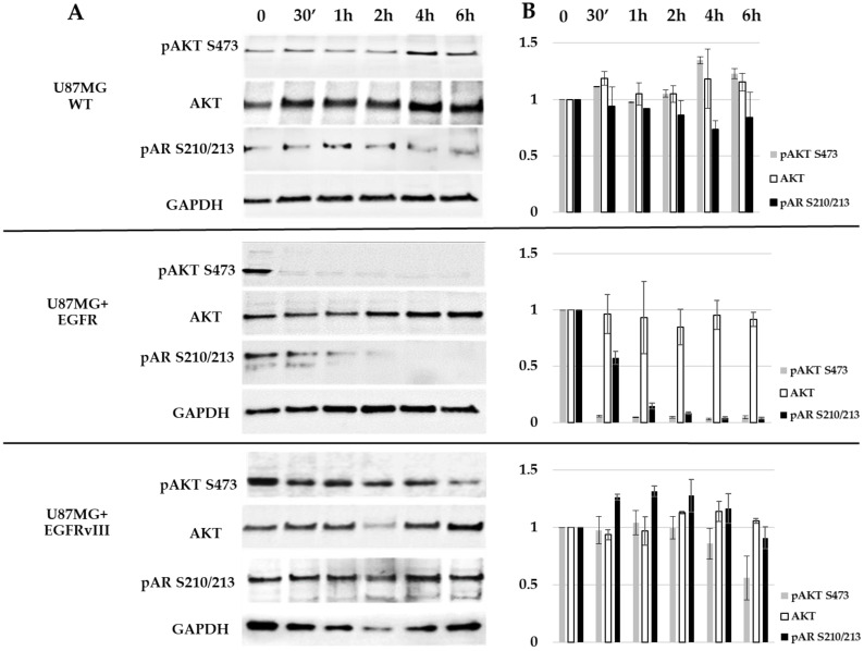
Figure 5.
Western blot analysis of AR S210/213 and AKT S473 in U87MG subclones. (A) Cells were treated for 30 min (30′) to 6 h with 5 μM afatinib and analyzed with Western blotting using antibodies against AKT, pAKT (S473), and pAR (S210/213). The anti-GAPDH housekeeping protein was used as a loading control. (B) Protein fold change (Y-axis) of each sample compared to treatment at time zero was calculated according to band densitometry analysis with ImageJ software, following normalization to GAPDH.

