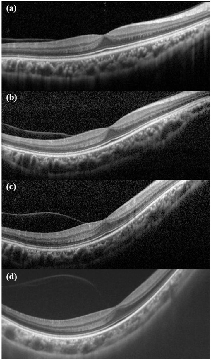
Figure 2.
Chronological changes of early PVD. (a) First OCT exam at age 7. At that time, the refractive error was −10 diopter. (b) After 3 years, it can be seen that PVD has partially progressed. (c) At the age of 13, the refractive error was −11.75D. Compared with (a), it can be seen that the contour of eyeball is more myopic. (d) At the age of 14, PVD showed further progress. Retinal detachment occurred 9 months later.

