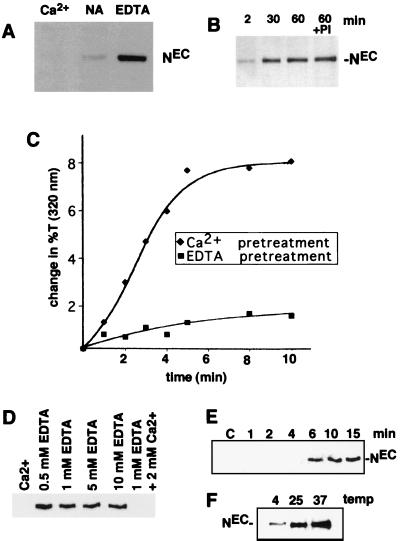
FIG. 4.
EDTA-induced dissociation of NEC from cells. (A) A total of 2 × 10⁶ N-S2 cells were incubated for 30 min at 25°C in TBS buffer containing 5 mM CaCl2 (Ca2+), no additional ions (NA), or 5 mM EDTA. Polypeptides within the conditioned media were then resolved by SDS-PAGE on 7% gels and transferred to nitrocellulose. A Western blot is shown that is stained with antibody F461.3B directed against the dN extracellular domain. (B) Kc cells (107) were incubated for various times at 25°C in TBS containing 0.5 mM EDTA. In one experiment, cells were incubated for 60 min in TBS containing 0.5 mM EDTA and a cocktail of protease inhibitors (PI) consisting of 1 mM Preflabloc (Roche), 10 μg of aprotinin per ml, and 10 μg of leupeptin per ml. (C) The effect of divalent metal ion chelation on the depletion of NEC from N-S2 cells was examined by a quantitative aggregation assay (see Materials and Methods). N-S2 cells were pretreated for 30 min at 25°C in 2 mM EDTA (■) or 5 mM CaCl2 (⧫). A molar excess of CaCl2 (5 mM) was then added to the EDTA-treated cells, which were incubated at additional 5 min. Dl-S2 cells were then added to Ca2+- or EDTA pretreated cells at 25°C. Aggregation was monitored by the change in transmitted light at 320 nm. (D) The concentration dependence of EDTA-mediated NEC shedding was investigated by incubating ∼2 × 10⁵ N1HA cells for 15 min at 37°C in HBS containing the indicated concentrations of CaCl2 and/or EDTA. Released NEC was precipitated from the conditioned media with anti-HA on protein A-beads, resolved by SDS-PAGE in a 6% gel, and detected on a Western blot stained with anti-HA. (E) The time course of NEC dissociation from N1HA cells was determined by incubating ∼2 × 10⁵ cells at 37°C in HBS containing either 2 mM CaCl2 (C) for 15 min or 0.5 mM EDTA for the times indicated. (F) The temperature dependence of NEC dissociation from N1HA cells was determined by incubating ∼2 × 10⁵ cells in TBS containing 0.5 mM EDTA for 15 min at the temperatures indicated. In panels E and F, the release of NEC into conditioned media was analyzed as described for panel D. Western blots stained with anti-HA are shown.

