Fig. 5.
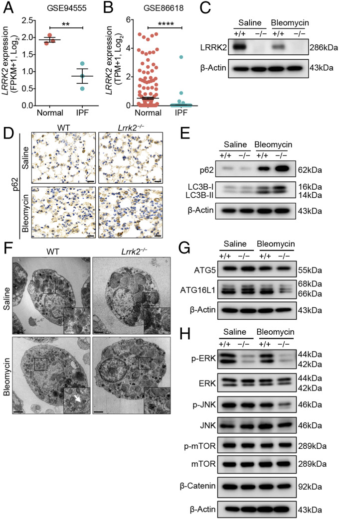
LRRK2 expression was down-regulated, and its deficiency led to impaired autophagy in AT2 cells in the progression of pulmonary fibrosis. (A and B) LRRK2 expression in AT2 cells from normal and IPF human lung tissues was determined based on published datasets (A) GSE94555 and (B) GSE86618 and displayed in dot plots. WT and Lrrk2−/− mice were treated with saline or 1.2 mg/kg bleomycin for 7 d (n = 3 to 4 mice per group). (C) Representative Western blot of LRRK2 expression in isolated AT2 cells. (D) Representative immunohistochemical analysis of p62 in lung sections. (Scale bars: 20 µm.) (E) Representative Western blot of p62, LC3B-I, and LC3B-II expression in isolated AT2 cells. (F) AT2 cells isolated from WT and LRRK2-deficient mice were evaluated by transmission electronic microscopy (original magnification: 4,200×). (Scale bars: 2 µm.) Magnified images of the boxed areas. The arrows indicate the autophagic vacuoles formed in bleomycin-treated WT mice. (G) Representative Western blot of ATG5 and ATG16L1 expression in isolated AT2 cells. (H) Representative Western blot of p-ERK, ERK, p-JNK, JNK, p-mTOR, mTOR, and β-Catenin expression in isolated AT2 cells. Three independent experiments were performed, and β-Actin was used as the loading control. Data are shown as mean ± SEM; **P < 0.01, ****P < 0.0001, by two-tailed Student’s t test (A and B).

