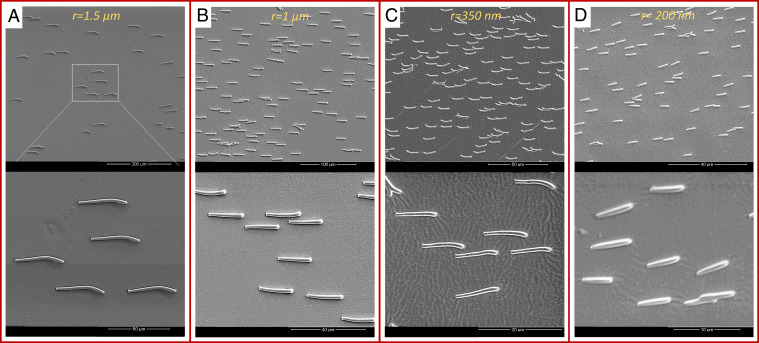
Fig. 2.
Scanning electron microscopy images of micro- and nanocilia. (A) Microcilia with , , and density . Images are made at different magnifications. (B) Microcilia with , , and density . (C) Nanocilia with , , and density . (D) Nanocilia with , , and density . All the cilia are fabricated and aligned in one direction by applying an external magnetic field while drying them in the ambient conditions.

